Menu
Your Cart

Leica DM6000 Upright Fluorescence Motorized Microscope with Motorized XY Stage (New Filters) Pred DM6

Leica DM6000 Upright Fluorescence Motorized Microscope with Motorized XY Stage (New Filters) Pred DM6

Most Microscopes ship within 3 to 10 business days

Hundreds of Custom Microscopes Shipped

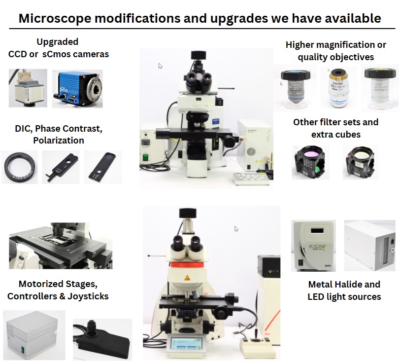
Customization available such as objectives, filter cubes, light sources, stages, cameras, and other components
Happy to provide references of past customers that bought microscopes from us

Save at least 60%+ vs. buying new and use those funds on another instrument instead.


  • Stock: In Stock
  • Brand: Leica
  • Product Code: SM100129-MG1MHXY
  • Condition: Pre-Owned
$9,995.00
CLICKABLE TABS BELOW

For Sale Leica DM6000 Upright Fluorescence Microscope Pred Leica DM6


Configured with a motorized XY stage suitable for petri dishes and slides.

Can be setup with other motorized stages  (microplate or multi-slide).


The Predecessor to Leica DM6 Upright Fluorescence Microscope


Condition, availability, and customization

  • Fully functional, serviced, and in good condition
  • Optics professionally cleaned 
  • *Comes with Brand New Fluorescence Filter Sets*
  • Can be custom configured to the requirements of your application
  • Will arrive timely, complete, and ready to be used


Leica DM6000 Fluorescence Microscope Overview 

(Final microscope capabilities will depend on the modifications you request)

Grade:

  • Research Grade Microscope

Microscopy types as configured:

  • For Brightfield and Fluorescence Microscopy

Suited for the following applications as configured:

  • High-quality imaging of live cells, tissues, microorganisms, and other

Installed automation/motorized components:

  • Motorized lightpath
  • Motorized/automated Z-Axis Drive
  • Motorized filter cube turret
  • Motorized objective(nosepiece) turret

Benefits of motorized/Automated Z-Axis drive:

  • Automatic parfocality adjustment
  • The ability to recall previously saved focus levels
  • Automated management of Z positioning/ Fine-coarse control

Imaging of Samples:

  • Ability to take still images and live videos of your samples

Imaging software highlights:

  • Display, annotate, edit, calibrate, measure, analyze, process, print, and save raster
  • Variable linear measurements such as line segment, radius values, and others 

Open source Micromanager Multi-dimensional Image acquisition & automation software capabilities

  • Multi-frame (Time lapse/Image acquisition over time) 
  • Multi-slice (Z coordinate/Z Stacking)   
  • *Multi-position (XY coordinate/Stitching/Mosaic) – only available when setup with motorized XY Stage


This is our Standard Leica DM6000 Fluorescence Microscope Configuration: 

Microscope Base:

  • Leica DM6000 upright Base
  • with a Motorized(Automated) Z-Axis Drive

Microscope Head:

  • Trinocular head with camera port
  • Two Eyepieces

Fluorescence Light Path:

  • Leica fluorescence motorized lightpath

Fluorescence Illumination:

  • EXFO X-Cite 120 Metal Halide source
  • Light Light Guide 
  • Collimating adapter  

Fluorescence filter cube turret:

  • Motorized 8-position filter cube turret

Fluorescence filters:

  • Three empty filter cubes
  • Three Brand New filter sets of either DAPI, FITC, Texas Red, or CY3

Brightfield Light Path:

  • Built into microscope base

Brightfield Illumination:

  • Leica 100 Watt halogen light source
  • Leica 100 Watt halogen power supply

Objective Turret:

  • Motorized 7-position objectives turret

Objectives:

  • 4x/0.10 PL objective 

  • 10x/0.30 PL Fluotar objective 

  • 20x/0.50 PL Fluotar objective

Condenser:

  • Mechanical condenser

Stage:

  • Motorized XY Stage 
  • Controller
  • Joystick

Stage Insert/Medium:

  • As configured may be used with slides or Petri dishes

(Motorized XY stage will be as shown or similar, guranteed to work with your sample)

Camera ports:

  • 100% Eyepiece, 0% Camera
  • 20% Eyepiece, 80% Camera
  • 0% Eyepiece, 100% Camera

Camera adapter:

  • 1x C-mount adapter 

Camera:

  • CMOS USB 3.0 10MP Camera

Computer:

  • Dell laptop computer

Software

  • Windows 10
  • Micromanager w/ImageJ Software
  • Camera software 

Accessories:

  • Necessary power and communication cables


Modifications and Upgrades Available (thousands of components on hand, priced on average 55-75% off the original price)

Most customers do elect to have microscopes configured to their requirements.

Configurations (In addition to installed Brightfield, and Fluorescence)

  • Phase Contrast, DIC, or Polarization Add-on Components may be added (subject to availability)

Maximum Magnification: 

  • Microscope can be configured for magnification between 40x and 1000x (with 10x Eyepieces)
  • Higher magnification Leica microscope objectives 
  • Various high-end objectives (APO) 

Fluorescence Capabilities: 

  • New filter set - DAPI
    (EX: 365/28nm, DM: 405nm, EM: 445/50nm)
    (Dye compatibility: DAPI/Hoechst/AlexaFluor 350)


  • New filter set - EGFP/FITC/Cy2
    (EX: 475/30nm, DM: 500nm, EM: 530/40nm)
    (Dye compatibility: EGFP/FITC/Cy2/AlexaFluor 488)


  • New filter set - TRITC/Cy3/Dsred
    (EX: 545/20nm, DM: 562nm, EM: 605/60nm)
    (Dye compatibility: TRITC/Cy3/RFP/Rhodamine/AlexaFluor 546)


  • New filter set - Texas Red
    (EX: 560/20nm, DM: 585nm, EM: 630/40nm)
    (Dye compatibility: Texas Red/mCherry/AlexaFluor 594)


  • New filter set - Cy5 (May require an upgraded light source) 

    (EX: 630/50nm, DM: 662nm, EM: 695/50nm)

    (Dye compatibility: Cy5/AlexaFluor 647/Draq5)


  • Extra Filter cubes


  • Xenon, Metal Halide, or LED Fluorescence microscope light sources

Imaging Capabilities: 

  • Various advanced CCD and sCMOS camera options for higher-quality images

Motorized/Automation Capabilities: 

  • In addition to installed motorized capabilities, it can be configured with:
  • Motorized high-speed shutter, motorized condenser

Biological Samples:

  • May be set up with other motorized XY stage kits for multi-slide scanning or full microplate scan


Happy to help build and configure the right microscope for you!


Most Requested Customer Upgrades

  • High-End CCD, sCMOS, or EMCCD Camera
  • LED Fluorescence Light Source
  • Phase Contrast, Polarization, or the DIC configurations
  • Higher Magnification Objectives
  • Higher performance Fluor or APO objectives
  • Motorized XY Stage Kit (stage, controller, joystick)


How to decide between a refurbished and brand-new comparable microscope

Will I save much by buying it pre-owned?

  • Add many upgrades noted above 
  • Add a 1-Year Extended Warranty
  • Add Setup and Training
  • Get imaging capabilities comparable to Leica DM6 Microscope
  • Get imaging quality comparable to Leica DM6 Microscope
  • Get all upgrades from one place
  • Still, save up to $60,000 vs buying new


Our Advantage

  • We are passionate about microscopy!
  • We offer detailed photos and video of the microscope in action
  • Are an approved vendor to most large US and Canadian Universities & Colleges
  • Are an approved vendor to many US and International Biopharma companies 
  • We carry hundreds of advanced research microscopes in stock 
  • Thousands of upgrade, modification, and repair parts in stock
  • Remote installation and microscope operation training packages
  • International shipping is no problem


Our Guarantee

  • In Stock, Multiple Leica DM6000 Microscopes on hand if you require more than one
  • The microscope comes with parts and a labor 90 Days Warranty
  • An extended 1-year warranty option may be available with optional yearly renewal for North American customers 
  • Remote installation and training options
  • Smooth process from the initial quote to custom order fulfillment
  • Customer references at many universities


Brochure: Leica DM6000 Upright Fluorescence Motorized Microscope

Manual: Leica DM6000 Upright Fluorescence Motorized Microscope


COMMON QUESTIONS AND ANSWERS BELOW


Steps you take to verify, service, and confirm the functionality of the Leica DM6000 Fluorescence Microscope?

Summary of testing & maintenance we perform (depending on included configuration):

  • Leica DM6000 upright Internal and external optics are inspected and cleaned
  • Mechanical components inspected, adjusted, and lubricated if necessary
  • The proper operation of electronic components is confirmed
  • Any necessary repairs and services completed to make sure the microscope is fully functional
  • Motorized components inspected for worn components, serviced and lubricated if necessary
  • Objectives are inspected to ensure there is no inside dust and a clear image is produced
  • Halogen lamp (if installed) we install a new Halogen bulb 
  • Fluorescence HBO Lamp (if installed) we install a new HBO bulb 
  • If the microscope has a Metal Halide Lamp source, we make sure the bulb has a minimum of 600 hours left on it
  • We inspect image quality of fluorescence images by looking at various fluorescing slide samples
  • Cameras are tested to ensure there is no condensation and they produce a clear image
  • Camera image quality verified

Can you help me identify the best microscope and configuration for me?

We are here to help

  • Contact us with your application; we will help you identify the best microscope configuration.
  • We do not believe in one size fits all approach; you should get a microscope that will do what you need.

Can I view the microscope in person at your facility or do a live Zoom demo before it is shipped to me?

We offer both onsite and remote demos via Zoom

  • We invite you to come in for an onsite or Zoom demo of the microscope
  • On a custom configured microscopes, we generally offer a demo before the microscope is ready to ship out

 What about the warranty?

Our Leica DM6000 upright Fluorescence microscopes come with a 90 Day Warranty.
 

Warranty Terms:

  • The warranty term is 90 Days from the date of the microscope's arrival at the destination.
  • This includes parts and labor.
  • The customer should retain all packing materials during the warranty period. 


Exceptions: 

  • The warranty does not cover bulbs except LED (LED lamps are covered)
  • Damage to the objectives or other optical components due to collision with a sample (improper use)
  • Electrical damage to the motorized/electronic components if the microscope plugged into an incorrect voltage


 Do you offer installation, service, etc?
  • We offer several remote installation and training packages.
  • We offer an extended warranty agreement on this model.
  • We stock various replacement and upgrade parts, so we may continue to support your microscope.


What should I watch out for when buying a Leica DM6000 Fluorescence Microscope used/pre-owned from another lab?

Basic steps to confirm quality:

  • Confirm that internal optics are clean; this is often a complex undertaking, especially complex microscopes with motorized components.
  • Confirm there is no present physical damage to the microscope base
  • Confirm there are no cracked lenses inside the microscope light paths or the base
  • Make sure there are no loose/damaged/seized gears inside mechanical and especially motorized components
  • Make sure a good amount of life left on fluorescence sources and light guides (if present) are damage free 
  • If Microscope has a motorized stage, make sure it does not skip steps and moves freely on all axis
  • Make sure objectives are installed, have been inspected, and have no damage or dust inside
  • Confirm that microscope is configured to your application; any changes later will be expensive
  • Confirm that camera is tested and communicates with the computer producing clear images
  • Make sure there are no missing components; it won`t be fast or inexpensive to correct


If you have concerns, we highly recommend either inspecting the microscope in person or doing a live Zoom call; this will allow you to evaluate the microscope and confirm that everything works on it.


 Do you have these and other microscopes on hand?
  • Subject to ongoing demand, we usually stock 100 to 150 of ready to go fluorescence microscopes (preconfigured)
  • Generally, we also have 150 to 200 microscopes in the process; those will be configured per the customer’s requirements.
  • We specialize in microscopes from Leica, Zeiss, Nikon, and Olympus.


This procurement is for a University or College, are purchase orders with NET terms accepted?
  • If you are a US or Canadian customer very likely we are already a registered and approved vendor in your University's vendor system(that is how many instruments we sold), this will simplify and expedite the process of PO approval.
  • POs are accepted from US and Canadian universities.


 How fast will you ship?
  • We can ship within a few days to 2 weeks, depending on your required Leica DM6000 microscope customization.


Will my instrument be insured during shipping?
  • All Leica DM6000 Fluorescence Microscopes are shipped fully insured within the US and internationally as long as we are a shipper and handle shipping arrangements.
  • If the buyer handles shipping, then shipping insurance responsibility falls to the buyer; we highly advise that the buyer obtain shipping insurance from their carrier.


 How do you pack? Have you packed and shipped microscopes in the past? Do you ship Internationally?

Packing

  • Most microscopes ship in fully enclosed ISPM-15 certified wooden crates, with proper padding underneath and the sides, and have moving components braced.
  • Accessories are packed safely in boxes with padding and then safely packed inside the crate.
  • We may not use a crate for local deliveries or smaller microscopes to save you money *if delivered by us.
  • On the crates, we often use “tilt’ & “shock” indicators to ensure the crate has not been excessively tilted and dropped where equipment may suffer damage.
  • Photos are taken during the packing process to make sure nothing is missed

USA/Canada Shipping

  • Within US and Canada, we will ship via selected freight carriers. We do not use low-price/high-damage carriers as we want the equipment to get safely to you.

International Shipping

  • Internationally we will only ship via Air Freight or Air Parcel, which provides reliability, no exposure to Ocean weather, and timely delivery.
    We have extensive experience shipping microscopes around the world.

Our experience and process

  • We have safely packed and shipped hundreds of complex customized microscopes.
  • Before packing/shipping, the instrument is re-tested, and a procedure is followed to verify the accuracy of included components.


How much will I save by buying it pre-owned, and is it worth it?

A lot…and it is certainly worth it, and here is why.  Feel free to ask for feedback from hundreds of customers that bought custom microscopes from us.

  • You will save around 60-80% vs. buying a new microscope.  Spend leftover funds on another instrument for your lab.
  • You will get a 90 days warranty, plus, when available, an option to get an extended warranty agreement. 
  • Many customers choose to invest funds into higher-end cameras that may not otherwise be considered due to cost at new prices.
  • Consider getting a motorized stage, often a $10,000 to $20,000 upgrade, when bought new. Often save over 70% on the stage alone.
  • For many applications, the difference in image quality between the current Leica DM6 microscope and this DM6000 may not make a major difference; instead, get better objectives or camera and have image quality exceed your expectations.
  • You can have Leica DM6000 upright microscope configured to provide excellent viewing/imaging quality for a fraction of the cost


Leica DM6000 Upright Fluorescence Microscope is a predecessor model to the Leica DM6 Fluorescence microscope; DM6 could be priced up to $95,000. So why not save your funds by buying Leica DM6000, get the high-quality image quality you expect from Leica, and use your leftover funds elsewhere?
 

 Sustainability
  • Well, this one is easy; buying a pre-owned Leica DM6000 Microscope is great for the planet; you are reducing unnecessary waste, CO2 output, and water use that would be used during the manufacturing process.  

 I have more questions; I want to receive a formal quote.

Absolutely! Please give us a call (1-617-366-2699) or complete this form to get your questions answered or receive a formal Quote.


Examples of past Leica DM6000 Microscopes that we built


Customer: Hvidovre Hospital, Denmark

SKU-32263


Customer Application: 

This customer was looking for a fully motorized fluorescence imaging microscope to view their cancer research. The Leica DM6000 is a modular microscope system, that allows customization and flexibility to meet the specific needs of cancer research.


Customer Requested Modifications:

  • From a full spectrum metal halide fluorescence source to a multi-band LED light source. LED light sources emit less heat and negligible amounts of UV or infrared radiation, minimizing phototoxic effects on live cancer cells during imaging. This is especially advantageous for long-term imaging studies or investigations involving sensitive cellular processes or drug response evaluations.
  • From our default DAPI, FITC, Texas Red filter sets to Attenuated DAPI, FITC, Texas Red, and CY3 filters. The Leica DM6000 can be equipped with fluorescence modules, such as fluorescence filter sets and fluorescence illuminators, allowing for precise fluorescence imaging and analysis of cancer cells and fluorescently tagged biomarkers.
  • The total magnification range of this microscope with 10x eyepieces and installed objectives was increased to 400x. The Leica DM6000 is known for its exceptional optical performance, providing high-resolution and contrast imaging. Its high-quality lenses and optics ensure clear visualization of cellular structures, which is crucial for studying cancer cells and their characteristics.
  • The stage was swapped from a manual XY stage to a motorized stage. A precise and stable stage with fine focus adjustment allows for accurate positioning and focusing of the slide containing cancer cells.
  • In combination with the base’s motorized Z focus, the new stage allowed for imaging options like Z stacking with mosaic stitching. The Leica DM6000 offers imaging automation capabilities, such as motorized stages, autofocus systems, and advanced software, enabling efficient and precise imaging of multiple samples, time-lapse experiments, and image analysis workflows.

Customer: Weill Cornell Medicine, New York

SKU-25856


Customer Application: 

The customer was looking for a way to scan a single slide for fluorescence immunology samples and requested phase contrast options. Fluorescence microscopy enables the detection and analysis of immune responses, including immune cell interactions, antibody labeling, and cytokine expression within tissues.


Customer Requested Modifications:

  • From our default, DAPI, FITC, Texas Red filter sets to FITC, Texas Red, and CY3 filtersImmunology often involves the use of fluorescent labeling to visualize specific immune cells, antibodies, or molecular markers. The Leica DM6000 can be equipped with fluorescence modules and filter sets, allowing precise and high-quality fluorescence imaging for immunological studies. 
  • Phase contrast annuals and matching objectives were added to allow for contrast imaging. Immunology studies often require visualization of cellular morphology and structural details. The Leica DM6000 supports various contrast techniques, including brightfield, phase contrast, and differential interference contrast (DIC), enabling researchers to observe immune cells and their interactions with enhanced contrast and detail. 
  • The total magnification range of this microscope with 10x eyepieces and installed objectives was increased to 630x. The Leica DM6000 delivers exceptional optical performance, including high-resolution imaging capabilities. This is critical for immunology research, as it allows researchers to observe and analyze fine details of immune cells, subcellular structures, or cellular interactions.
  • The CMOS camera was upgraded to a CCD which delivers exceptional optical performance, including high-resolution imaging. This is critical for immunology research, as it allows researchers to observe and analyze fine details of immune cells, subcellular structures, or cellular interactions.
  • The new stage allowed for full slide scanning using image capture and processing software. These automation capabilities enhance workflow efficiency and enable high-throughput imaging and data acquisition.  

Customer: International Independent Researcher

SKU-24565


Customer Application: 

This customer was looking for a reasonably priced time-lapse slide-imaging microscope for biological studies with DIC options for Pharmacological research. The Leica DM6000 microscope provides valuable functionality and performance for pharmacology and drug discovery research. It empowers researchers to visualize, analyze, and understand the effects of drugs on cellular processes, contributing to the development of new therapeutics and advancements in pharmacological science.


Customer Requested Modifications:

  • From our default, DAPI, FITC, Texas Red filter sets to DAPI, FITC, Texas Red, and CY3 filters. Pharmacology and drug discovery often involve the use of fluorescent probes, dyes, or markers to study drug-target interactions, cellular responses, and drug efficacy. The Leica DM6000 can be equipped with fluorescence modules and filter sets, enabling precise and high-quality fluorescence imaging for pharmacological studies. 
  • Differential Interference Contrast prisms and matching objectives were added to allow for contrast imaging. The Leica DM6000 supports various contrast techniques, such as brightfield, phase contrast, and differential interference contrast (DIC). These techniques facilitate the observation of cellular morphology, structural changes, and drug-induced alterations, enhancing the visualization of cellular responses to pharmacological treatments. 
  • The total magnification range of this microscope with 10x eyepieces and installed objectives was increased to 400x. The Leica DM6000 delivers exceptional optical performance, providing high-resolution imaging capabilities. This is crucial in pharmacology and drug discovery research to observe and analyze cellular structures, drug uptake, intracellular localization, and drug-target interactions at a microscopic level.
  • The stage was swapped from a manual XY stage to a motorized stage. Pharmacological studies often require imaging multiple samples or conducting high-throughput experiments. The Leica DM6000 features motorized stage and imaging automation capabilities, enabling efficient and precise sample positioning, multi-site imaging, and time-lapse experiments. Automation reduces human error and enhances workflow efficiency.
  • The head was swapped for a more ergonomic model. Prolonged microscope use is common in pharmacology research. The Leica DM6000 is designed with ergonomic considerations, such as adjustable eyepieces, comfortable stage controls, and customizable settings, ensuring user comfort during extended periods of observation, analysis, and experimentation. 

Customer: Research Facility in Shanghai China 

SKU-30483
Customer Application: 

This customer was looking for a budget-friendly motorized microscope for viewing cells for neurological research. In neuroscience, fluorescence microscopy helps researchers visualize neuronal structures, track neural activity, study synaptic connections, and investigate the mechanisms underlying neurological disorders.


Customer Requested Modifications:

  • Our default DAPI, FITC, and Texas Red filter sets were changed to Texas Red, CY3, and CY5. Neurological studies often require the simultaneous imaging of multiple fluorophores or fluorescent labels to investigate complex neuronal processes or multiple molecular targets. The Leica DM6000 supports multi-channel imaging, allowing researchers to capture and analyze multiple fluorescent signals within a single sample.
  • The total magnification range of this microscope with 10x eyepieces and installed objectives was increased to 630x. The Leica DM6000 provides excellent optical performance, including high-resolution imaging capabilities. This is crucial in neurology research to observe and analyze fine details of neuronal structures, synaptic connections, and cellular morphology.
  • The addition of Micromanager and FIJI software allowed for quantitative analysis of neuronal structures, such as dendritic spine density, axonal length, or neuronal connectivity. The Leica DM6000 combined with imaging software for automated image analysis, data quantification, and statistical analysis, facilitating accurate and efficient data processing.
  • The stage was swapped from a manual XY stage to a motorized stage. The Leica DM6000 offers imaging automation features, including motorized stages, which are valuable for high-throughput imaging, multi-site imaging, and time-lapse experiments in neurology research. Automation enhances workflow efficiency and allows for precise and reproducible imaging.
  • The new stage allowed for joystick control which allowed for quick scanning of their samples during viewing. Neurology research can involve long hours of microscope use. The Leica DM6000 is designed with ergonomic considerations, such as adjustable eyepieces, comfortable stage controls, and customizable settings, ensuring user comfort during extended periods of observation and analysis.

Customer: Baylor University, Texas

SKU-26413


Customer Application: 

This customer requested a fully motorized microscope that could be used for studying the development of organisms by visualizing morphological changes, tracking cell migration, and examining gene expression patterns during embryogenesis.


Customer Requested Modifications: 

  • From our default, DAPI, FITC, Texas Red filter sets to DAPI, FITC, Texas Red, and CY3 filters. Developmental biology often involves the use of fluorescent markers or dyes to visualize specific developmental processes, gene expression patterns, or cell lineage tracing. The Leica DM6000 can be equipped with fluorescence modules and filter sets, enabling precise and high-quality fluorescence imaging for studying cellular dynamics and molecular interactions during development. 
  • The total magnification range of this microscope with 10x eyepieces and installed objectives was increased to 400x. The Leica DM6000 provides exceptional optical performance, including high-resolution imaging capabilities. This is crucial in developmental biology to observe and analyze fine details of embryonic structures, tissue morphogenesis, and cellular differentiation during various stages of development.
  • The stage was swapped from a manual XY stage to a motorized stage. The Leica DM6000 offers imaging automation features, including motorized stages and autofocus systems, which are valuable for high-throughput imaging, time-lapse experiments, and multi-site imaging in developmental biology research. Automation enhances workflow efficiency and allows for precise and reproducible imaging.
  • The new stage allowed for a full slide scanning function to be added to time-lapse image capture. Utilizing Micromanager and FIJI software, quantitative analysis of developmental structures, such as cell counts, tissue measurements, or growth dynamics is possible. The Leica DM6000 combined with imaging software for automated image analysis, data quantification, and statistical analysis, facilitating accurate and efficient data processing.

Customer: Doyle Lab, Arizona 

SKU-15307


Customer Application: 

This customer wanted an upright microscope for viewing samples at very high magnification in molecular biological studies. The Leica DM6000 microscope provides researchers in molecular biology with the necessary tools and performance to investigate molecular interactions, cellular processes, and molecular mechanisms. It aids in advancing our understanding of gene expression, molecular signaling pathways, and molecular interactions, contributing to the field of molecular biology. 


Customer Requested Modifications:

  • From our default, DAPI, FITC, Texas Red filter sets to DAPI, Texas Red, and CY3 filters. Molecular biology often involves the use of fluorescent probes, dyes, or markers to visualize specific molecules, gene expression patterns, or cellular events. The Leica DM6000 can be equipped with fluorescence modules and filter sets, enabling precise and high-quality fluorescence imaging for studying molecular interactions, protein localization, and cellular processes. 
  • The total magnification range of this microscope with 10x eyepieces and installed objectives was increased to 1000x. The Leica DM6000 provides exceptional optical performance, including high-resolution imaging capabilities. This is crucial in molecular biology to visualize and analyze subcellular structures, cellular processes, and molecular interactions at a microscopic level.
  • APO objectives were selected for their superior imaging capabilities.APO objectives are designed to minimize chromatic aberration, which is the phenomenon that causes different colors of light to focus at slightly different points. This reduction in chromatic aberration improves image quality by enhancing color accuracy, contrast, and resolution. It allows for clearer and more precise imaging of samples, especially when using multiple wavelengths or fluorescence imaging.
  • The stage was swapped from a manual XY stage to a motorized stage. The Leica DM6000 offers imaging automation features, including motorized stages and autofocus systems, which are valuable for high-throughput imaging, time-lapse experiments, and multi-site imaging in molecular biology research. Automation enhances workflow efficiency and allows for precise and reproducible imaging. The new stage allowed for the still placement of the samples during oil immersion image capture
  • The addition of Micromanager and FIJI software allows for quantitative analysis of molecular structures, protein expression levels, or cellular dynamics. The Leica DM6000 can be combined with imaging software for automated image analysis, data quantification, and statistical analysis, facilitating accurate and efficient data processing.


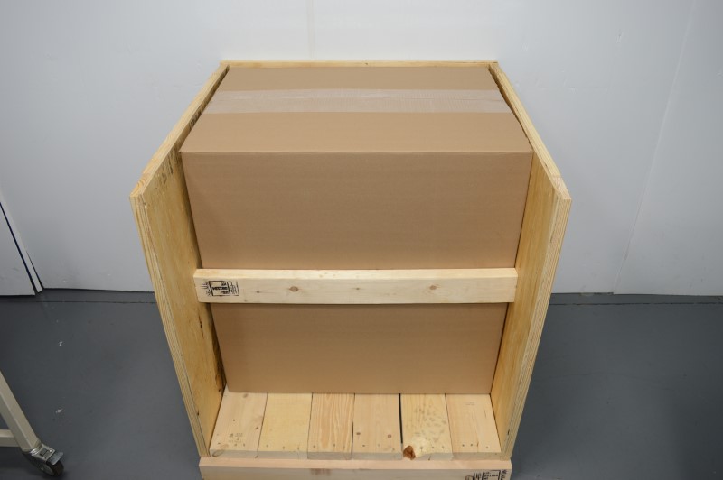
Freight Microscope Packaging Process

At our company we take packing and shipping very seriously.
No one wants to spend thousands of dollars and receive a damaged instrument.

Our process involves evaluating every microscope and its components to determine the safest positioning and structure for packing.
Most of our microscopes will ship in an ISPM-15 wood crate made from ½” plywood and 1x4 framing. (Crates are Internationally Compliant).

During microscope packing we often disconnect any protruding components like lamp housings, sliders, eyepieces, light guides.
We provide easy reassembly instructions to reinstall such components.

Components and accessories not installed on the microscope are bubble wrapped and packed in individual boxes.
Any remaining space inside the crates is filled with bubble wrap to prevent any movement and provide for additional cushion.

All of our crates ship with carrier instructions “Do Not Stack“ and include visual customer reminders to inspect the shipment on arrival.
Crates are also marked with instructions to retain packaging material until the instrument is inspected/verified and warranty period passed.


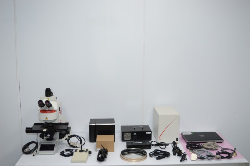
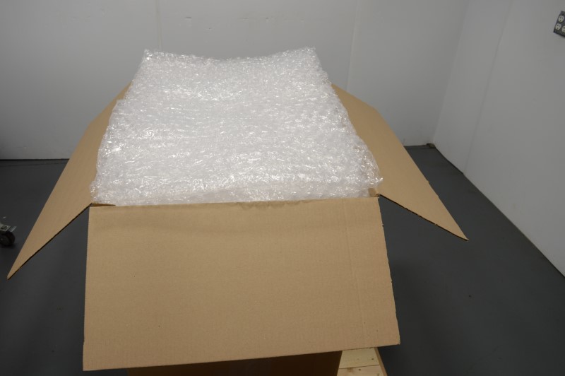
Parcel Microscope Packaging Process

Same as with freight shipments we take packing and shipping very seriously.
No one wants to spend thousands of dollars and receive a damaged instrument.

Our process includes evaluating every microscope and its components to determine the best shipment and packing method.

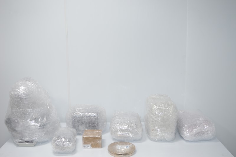
Microscopes that are small enough to ship via parcel (UPS/FEDEX) are often taken apart with all components safely wrapped with bubble wrap.
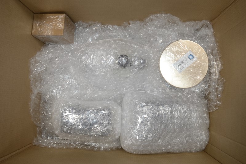
Safety of shipment is a number one priority so we may often pack microscopes into multiple boxes to assure safe delivery.

Boxes have a foam bottom liner and are filled with bubble wrap to prevent any movement of content inside during transit to your location.


Safe Shipping
From Small Packages to Large Freight
International Shipping
Happy customers all around the world
Warranty
60-days
Registered Vendor To
Many US Colleges, Universities and Fortune 500 Companies